Zusammenfassung
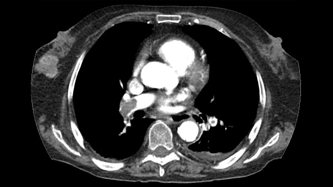
In der konventionellen Radiologie gehört der Thorax zu den am häufigsten untersuchten Regionen. Durch die Fortschritte bei Computertomografie und Magnetresonanztomografie konnte die Auflösung erheblich gesteigert werden. Das erhöht die Früherkennung von Krankheiten. Dabei ist es wichtig, immer auch ein Auge auf die mitabgebildeten Organe und Strukturen zu werfen. Eine Umgebungsdiagnostik ist bei der Anfertigung der CT-Bilder enorm wichtig. Je häufiger sich eine MTR am Gerät die angefertigten Bilder auch anschaut, desto schneller kann man auf eventuelle Zufallsbefunde reagieren und gegebenenfalls das Untersuchungsmanagement anpassen. Einige Fallbeispiele werden im Beitrag vorgestellt.
Schlüsselwörter: Thorax, Früherkennung, Zufallsbefunde, CT, MRT
Abstract
The thorax is one of the most frequently examined regions in conventional radiology. Thanks to advances in computed tomography and magnetic resonance imaging, the resolution has increased significantly. This increases the early detection of diseases. It is important to always keep an eye on the organs and structures that are also shown. Surrounding diagnostics are extremely important when preparing the CT images. The more often an MTR/radiographer looks at the images produced on the device, the faster one can react to any incidental findings and, if necessary, adapt the examination management. Some case studies are presented in the article.
Keywords: thorax, early detection, incidental findings, CT, MRI
DOI: 10.53180/MTIMDIALOG.2023.0344
Entnommen aus MT im Dialog 5/2023
Dann nutzen Sie jetzt unser Probe-Abonnement mit 3 Ausgaben zum Kennenlernpreis von € 19,90.
Jetzt Abonnent werden

